Microphysiological systems
Development of microphysiological systems, organ-on-chips and microfluidics for biology
Fabrication of an environmental-controlled human colon-like micro-physiological system
Doctoral researcher: Duvan Rojas Garcia
Associated Researchers : Marie Hut
Supervision: Laurent Malaquin / Julie Foncy (I2C) / Audrey Ferrand (IRSD)
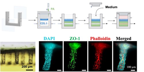
We develop human colon-like MPS, recapitulating faithfully in vitro the 3D human colonic topology and matrix support stiffness. These MPS integrate a microfluidic chamber that allows active control of both the apical/luminal and basal/stromal compartments, as well as in situ imaging. The system successfully supported colonic epithelial Caco-2 cells culture, reproducing human colon-crypts geometry and dimension. Epithelial constructs were highly polarized and expressed functional markers such as tide junction’s proteins and mucus secretion. Overall, this innovative MPS recapitulates cell microenvironment cues and offers a high observability, making it a useful tool to study the colon epithelium, its interaction with external factors and drug screening.
Funding : PEPR MEDOOC (https://www.pepr-medooc.fr/)
Development of intestinal microphysiological systems
Post-Doctoral researcher: Elise Ponthier
Supervision: Laurent Malaquin / Arnaud Besson (CBI)
Associated Researchers : Julie Foncy, Daniel Ferri Angulo, Lucien Guth
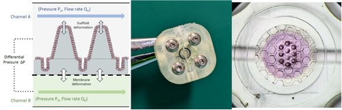
The microphysiological systems will recapitulate microenvironnemental parameters of in vivo organ such as topography, peristaltic movements and biochemical gradients. To do so, we are combining 3D printing for the topography and microfluidics to integrate mechanical cues. We developed a prototype microfluidic chip composed of two channels with a honeycomb membrane in between. We succeeded in 3D printing our intestinal scaffold directly onto this membrane. First tests are performed with Caco-2 and the next ones will be carried out with intestinal organoids. Based on this model, we aim to study intestinal stem cell fate and early steps of tumorigenic transformation.
Perfused skin in-vitro model
Researcher: Laurent Malaquin
Collaboration : Nicolas Giang & Emeline Pagès (Genoskin), Nicolas Gaudenzio (Infinity)
The primary function of the skin is to act as a barrier against external aggressions. Skin protection mechanisms rely partly on the presence of resident immune cells and the recruitment of circulating immune cells from the blood. Genoskin has developed two ex vivo human skin models: a perfused model using a porous catheter implanted in the tissue and a wound model. Additionally, a blood sample is collected and matched to each skin sample.
Based on three key resources—an implantable device, wounded skin, and blood—the project's goal is to model, ex vivo, the recruitment of circulating immune cells in a human skin wound model that closely mimics clinical conditions. This microfluidic platform is adaptable to other solid tissues and will enable the study of immune cell recruitment and infiltration in various inflammatory and pathological contexts.
Funding : ANR CIMMS
Modular microfluidic modules and frames for gut epithelium culture
Post-doctoral researcher: Matthieu Sagot
Supervisor: Bastien Venzac
Description of the project