
PEGDA 3D scaffolds reproducing intestinal epithelium topography
The small intestine is a complex tissue with a crypt/villus architecture and high tissue polarity. The maintenance of tissue integrity and function is supported by a constant renewal of the epithelium, with proliferative cells located in crypts and differentiated cells migrating upward to the top of villi. So far, most in vitrostudies have been limited to 2D surfaces or 3D organoid cultures that do not fully recapitulate the tissue3D architecture, microenvironment and cell compartmentalization found in vivo. Here, we report the development of a 3D model that reproduces more faithfully the architecture of the intestinal epithelium in vitro. We developed a new fabrication process combining a photopolymerizable hydrogel that supports the growth of intestinal cell lines with a high-resolution stereolithography 3D printing. This approach offers the possibility to create artificial 3D scaffolds matching the dimensions and architecture of mouse intestinal crypts and villi. We demonstrate that these 3D culture models promote the growth and differentiation of Caco-2 cells for 3 weeks. Interestingly, we observed a clear increase in cell polarization on 3D hydrogel scaffolds compared to 2D ones, suggesting that both material and topography promote cell differentiation. These models offer intriguing alternatives to organoid cultures to study tissue homeostasis or to perform in vitrodrug screening.
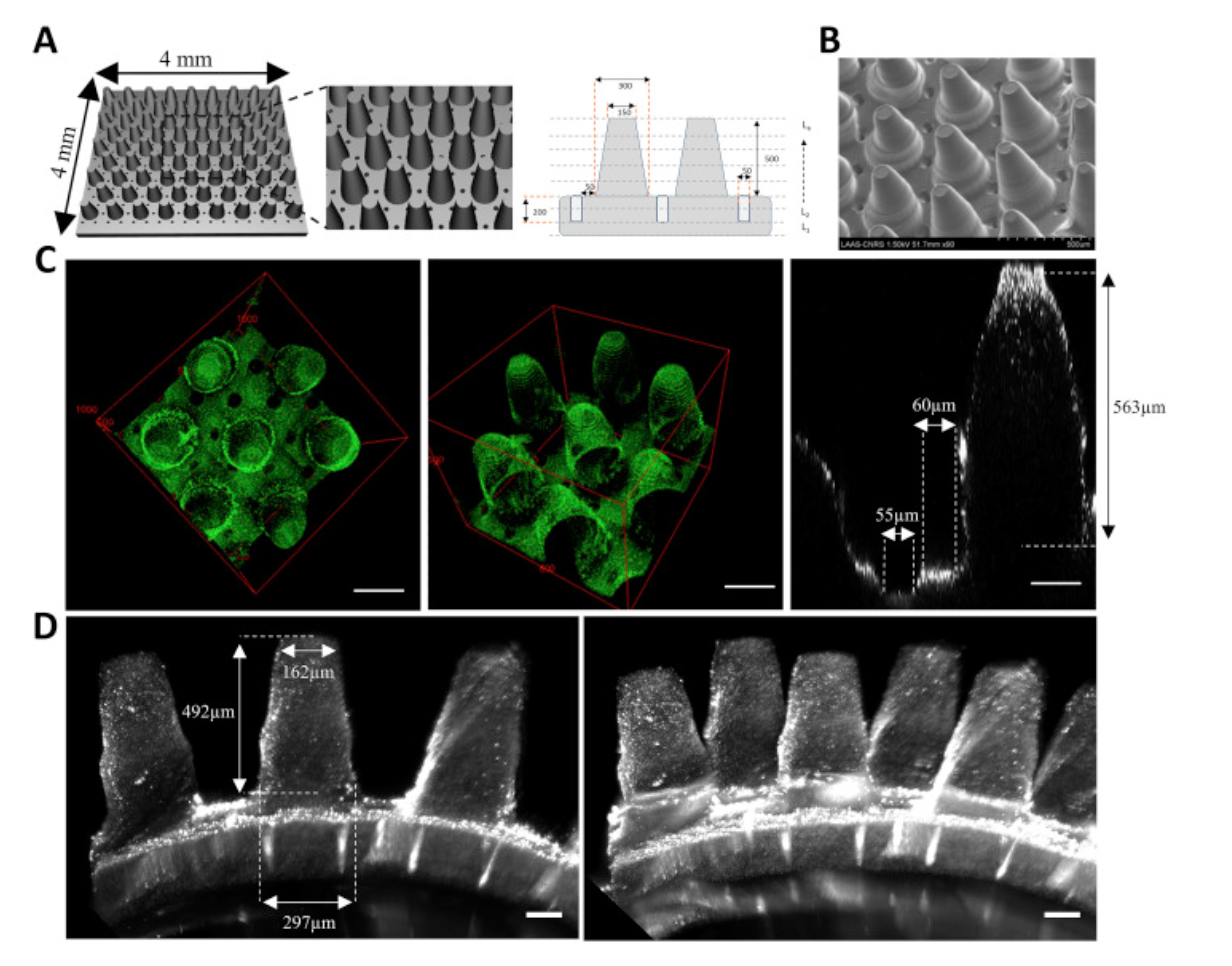
3D printing and imaging of intestinal scaffold. A. CAD design and schematic of the 3D intestinal scaffold mimicking the topography and dimensions of murine intestinal epithelium. Numbers are in μm. Scaffolds were printed using a photopolymerizable hydrogel composed of 40% PEG-DA 700, 30% acrylic acid and 250 μg/ml fibronectin, fluorescent beads were added to visualize the scaffolds by fluorescence microscopy. B. SEM images of the 3D scaffold. C. 3D reconstructions (left and middle) and xz section (right) of the scaffold image by two-photon microscopy. D. Side view of the scaffold image (left) with Light Sheet Fluorescence Microscopy and 3D reconstruction (right). Scale Bars = 100 μm.
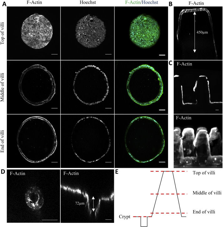
Cell culture on intestinal scaffolds. Representative images of Caco-2 cells seeded on intestinal scaffolds. Cells were grown for 6 days fixed and then stained for DNA (Hoechst 33342, blue) and F-actin (FITC-Phalloidin, green). A. Two-photon imaging of the top, middle and base of the villi. Scale bars = 50 μm B. xz section of a single villus imaged by two-photon microscopy. C. Side view of the scaffold (top) and 3D reconstruction (bottom) of Light Sheet Fluorescence Microscopy acquisition. Scale bars = 100 μm. D. xy and xz confocal microscopy images of the crypt compartment. Scale bars = 50 μm. E. Schematic showing the different horizontal cross-sections imaged on the 3D scaffold.