
3D hydrogel scaffolds for neuronal cell growth
3D fabrication techniques are rapidly expanding in the field of scaffold development for cell culture and tissue engineering. Herein we report the realization of free-standing PEGDA hydrogel architectures by using two-photon lithography. The morphological and immunofluorescence characterization of neuro2A cells revealed a tridimensional colonization featuring multiple neuritic extensions per cell as well as the expression of β-tubulin neuronal marker and actin microfilaments. The results open new perspectives in the continuous quest for structured biomaterials able to provide a favorable environment to cells and at the same time not interfering with imaging protocols necessary for a clear scenario of the cell seeding.
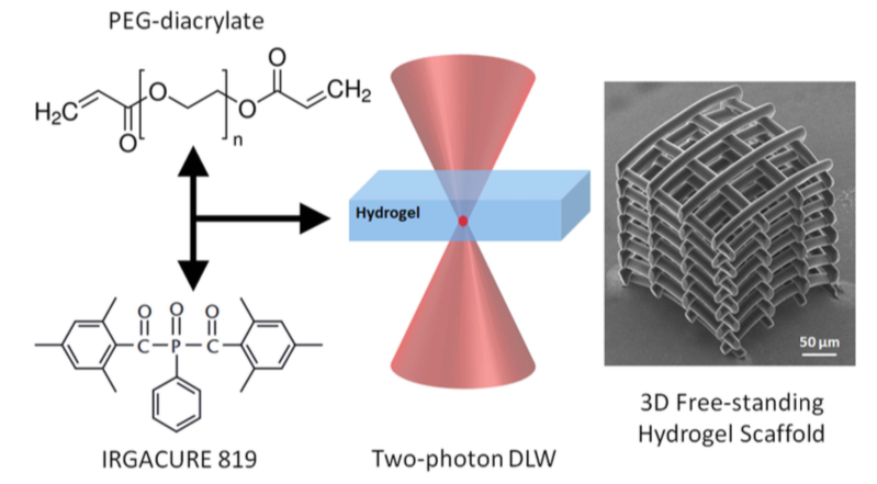
A: schematic design of the PEGDA solution preparation and 2PL-DLW fabrication of the 3D hydrogel scaffold.
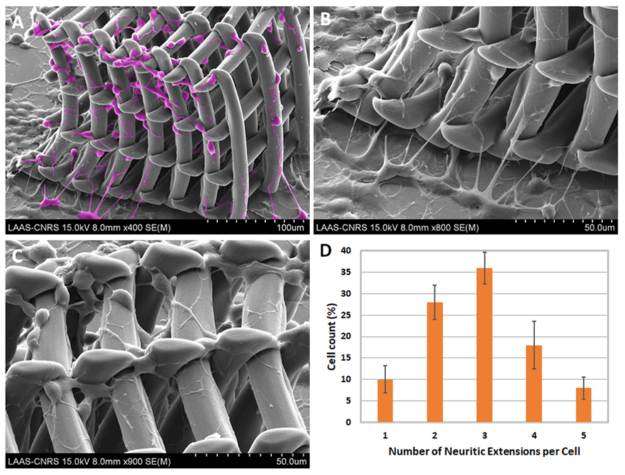
(A)–(C): scanning electron microscopy characterization of a 3D PEGDA hydrogel scaffold colonized by neuro2A cells ((A): false-colored SEM micrograph highlighting the colonization of neuro2A cells on the 3D PEGDA scaffolds. (D): histogram highlighting the number of neuritic extensions per cell (data are given as mean ± standard deviation).
